
.jpg) 1 hour ago
2
1 hour ago
2
The human brain is creatively malleable, producing breathtaking art such as The Starry Night (twice) and the Large Hadron Collider to reveal different sides of reality, including the bowling-pin-shaped atomic nucleus of neon-20.
The brain is physically malleable as well, showing plasticity that reshapes its neuronal connections, cell types, and signaling pathways.
These changes can result from neurological conditions, the as-yet-inevitable effects of aging, or, say, eating tomato paste for three months.
Brain plasticity can also occur in response to one's sensory experiences. For example, a variety of scientific literature has described brain changes and improved visual capacities in D/deaf persons.
A new brain-imaging study suggests that adults living with profound deafness since early childhood devote relatively more of their early visual system to the far edges of their field of view.
In their paper published in PNAS, a team of psychologists and neuroscientists in the UK showed that this 'reorganization' appears surprisingly early in the visual pathway, before signals even reach the brain's main vision center.
The effects could be important for detecting potential hazards.
"Without sound cues, deaf people use vision as an 'early warning system' for peripheral events," the researchers say.
"Indeed, D/deaf adults exhibit superior performance in peripheral tasks involving detection or attention to transient or moving targets."
The researchers recruited 16 adult participants with early, profound deafness and 16 hearing, age-matched individuals for the control group.
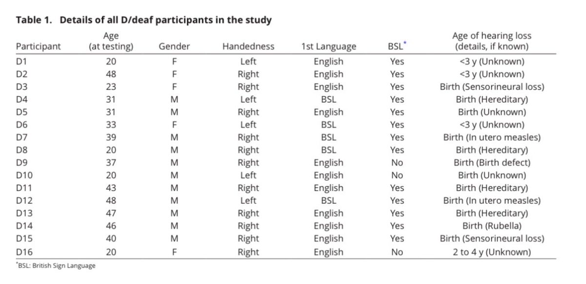 The study participants' details. (Levine et al., PNAS, 2026)
They used structural magnetic resonance imaging ( MRI) to analyze neural structure and functional MRI to perform retinotopic mapping. This technique maps which brain regions respond to different locations in a person's visual field.
The researchers focused on two key regions. These include the lateral geniculate nucleus (LGN) of the thalamus, which functions like a relay station for visual information arriving from the retina after traveling through the optic nerve.
The LGN projects to the primary visual cortex (V1), the major visual-information-processing region at the back of the brain.
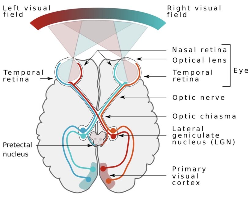 An illustration of the brain's visual systems. (Miquel Perelló Nieto/Wikimedia Commons/CC-BY 4.0)
The researchers' goal was to align brain activity with individuals' actual visual fields and compare differences between the two groups.
They did so by calculating the proportion of active voxels (3D MRI units representing small volumes of brain tissue) across one's visual field, by splitting said field into eccentricity "bins," or concentric slices of vision moving away from one's central focus.
They then linked brain activity to these slices of the visual field to analyze the relationships between brain tissues and vision – specifically, which visual brain regions "respond most strongly to which part of one's vision.
 The 'slices' of vision used to explore eccentricity preferences. (Levine et al., PNAS, 2026)
To ascertain that these results were not biased by how they defined brain boundaries, the researchers also compared their hand-drawn retinotopic boundaries for V1 with two standardized brain atlases, or maps, including one from the Human Connectome Project.
Altogether, the findings suggest that LGN activity is relatively biased toward peripheral vision in D/deaf persons compared with hearing persons, suggesting that visual reorganization in the brain starts at least as early as the LGN.
Yet, interestingly, the researchers found no significant difference in LGN volume between the two groups.
The neural bias for peripheral vision persisted as the researchers delved digitally deeper into the brain's visual processing systems to explore visual field representations in V1.
Again, they found no difference in total V1 volume between the two groups.
But they did find that the volume was distributed differently. D/deaf persons displayed neural preferences for peripheral vision, while hearing persons showed neural preferences for more central vision.
Notably, these volumetric differences appeared to reflect changes in cortical surface area rather than cortical thickness.
Altogether, the lifelong greater reliance on vision in people with early, profound deafness appears to be linked to compensatory plasticity in the LGN and V1 in response to underutilized auditory brain systems.
The study doesn't show that D/deaf people see better in every situation.
This leads to a visual trade-off, the researchers say, revealing "a redistribution of neural resources in early D/deaf individuals, with a larger cortical surface representation of the periphery, at a cost of smaller representations of the central visual field."
In an exploratory analysis too small for valid statistical testing, the largest increase in peripheral-vision preference, and the accompanying trade-off to central vision, was apparent in D/deaf individuals who learned British Sign Language (BSL) as their first language.

As per past evidence, the researchers say neural differences in D/deaf individuals could arise from an even more upstream source: the retina – light-sensitive, neuron-rich tissue at the back of the eye that turns light into electrical signals sent to the brain via the optic nerve.
Related: Chronic Tinnitus Isn't Just Ringing – It's Rewiring The Brain
Finally, the researchers note that some of the D/deaf persons included in this work were genetically deaf. In contrast, others had unknown causes underlying their deafness, which may suggest the influence of some unidentified genetic precursor.
Future studies could further explore the relationship between genetic deafness, visual enhancement, and specific brain plasticity processes.
This research was published in the Proceedings of the National Academy of Sciences.
This article was fact-checked by Rebecca Dyer and edited by Rebecca Dyer. While we pride ourselves on our process, we are only human. If you spot a mistake, please let us know.
 English (US)