
.jpg) 1 hour ago
4
1 hour ago
4
Up until the 1970s or so, the human brain was assumed to be 'hardwired' like a computer, its neural networks remaining fixed throughout adulthood.
In reality, this incredible organ can reinvent itself time and time again.
Like Taylor Swift's career, the human brain has several major eras – and motherhood seems to be no exception.
In 2024, neuroscientists in the US provided the first detailed map of the human brain over the course of a single pregnancy.
The woman who participated in the research allowed brain scans to be taken before, during, and after gestation.
As her due date approached, researchers noticed parts of her brain were shrinking in volume.
The metamorphosis was unlike anything scientists had ever seen.
That prompted the launch of the Maternal Brain Project (MBP): a research initiative to better understand how the brain changes during motherhood – a topic that experts have severely neglected.
Since 1990, only 0.5 percent of brain scan studies have looked at women's health, according to neuroscientist Emily Jacobs, who leads MBP at the University of California, Santa Barbara (UCSB).
Jacobs and her team are now taking the project global, with the project currently being expanded to include more institutes in the US and one in Spain.
Brain scans from several more mothers since 2024 have found the same widespread volume losses.
What's more, there seem to be changes in the brain's vascular system during pregnancy, including modifications in how cerebrospinal fluid flows.
"Nearly every region in the brain is changing significantly across pregnancy," Jacobs told ScienceAlert.
"We know that the cardiovascular system undergoes profound changes across pregnancy to accommodate the growing fetus, and from this data we are observing that the cerebrovascular system undergoes striking adaptations to pregnancy as well."
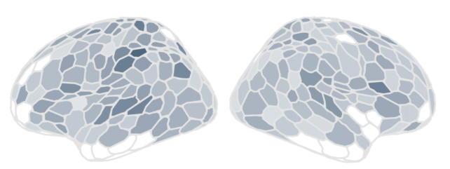 In the 2024 study, widespread cortical gray matter volume change occurred in step with advancing gestational week. Darker colors indicate regions most affected by the pregnancy transition. (Laura Pritschet)
The results are not yet published in a peer-reviewed journal, but of all 400 brain regions Jacobs and her colleagues have examined, she says 97 percent showed alterations across gestation and after birth.
A loss of total brain volume may sound like it would have negative effects, but in all likelihood, these sweeping changes reflect how the brain reshapes itself to become more efficient for motherhood.
After all, this highly malleable organ can only use a finite amount of tissue to deal with major life events, like puberty, pregnancy, or menopause.
"This project, and others like it, are updating the way we think and talk about matrescence," Jacobs said, "dispelling outdated notions of 'mommy brain' as being dysfunctional or inadequate, and instead portraying the maternal brain as one capable of continual brain adaptation and neural plasticity."
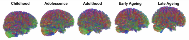 Some evidence suggests that the human brain features five eras throughout life, regardless of sex. (Gates Cambridge)
PhD student Hannah Grotzinger has led the Maternal Brain Project's ongoing data collection effort.
The study now includes 20 participants in the US, including first-time mothers, second-time mothers, and two comparison groups: fathers and non-pregnant women.
These participants were tracked on a regular basis over an 18-month-long period, using MRI scans, blood draws, and questionnaires on mood, sleep, and health.
So far, the team has conducted more than 150 scans, and among first-time mothers, Jacobs, Grotzinger, and colleagues are seeing highly similar structural changes.
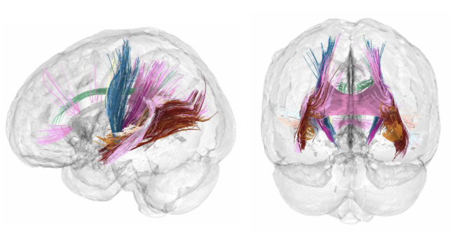 Major white matter tracts demonstrated increasing microstructural integrity over pregnancy. (Daniela Cossio)
The total volume of their brains seems to decrease linearly during pregnancy, before showing some signs of returning after the child is born.
"We are seeing broadly consistent patterns of structural remodeling throughout the brain," explained Jones.
"Total brain volume, gray matter volume, and cortical thickness decrease significantly across pregnancy, and partially rebound in the postpartum period, with cerebrospinal fluid following the opposite pattern."
What's more, these changes are occurring in the same brain areas as the very first participant. These include the superior temporal lobes, the midline, the prefrontal cortex, and subcortical regions.

The next step is to build a larger and more diverse cohort of mothers to see if individual factors, like fertility treatments, pregnancy complications, or breastfeeding, may influence how the brain rewires itself during pregnancy and in the postpartum period.
The goal is to establish the most comprehensive, open-access maternal brain database in the world.
That way, researchers can start answering questions like: How does remodeling of the maternal brain impact cognition? Can we detect early biological signs of postpartum depression?
And what lasting effects do pregnancy conditions like preeclampsia or gestational diabetes have on brain health?
Related: Your Brain Goes Through 5 Distinct Epochs, Massive Study Finds
Neuroscientists at USCB will help lead the research, along with scientists at the University of Pennsylvania and a research institute in Spain called IISGM.
In 2023, Jones wrote a perspective article for Nature highlighting major sex disparities in brain health research.
"Representation is not the problem: about 50 percent of people enrolled in neuroimaging studies listed on OpenNeuro.org are women," she wrote.
"Researchers are simply not choosing to study (and funders to invest in the study of) health factors specific to women."
The Global Maternal Brain Project is on a mission to change that.
 English (US)